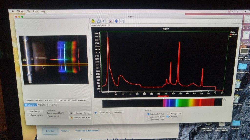
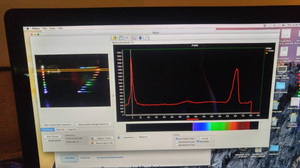
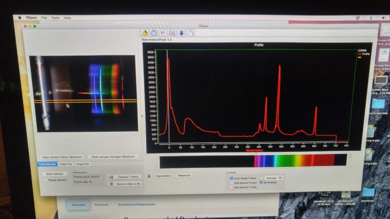
- The Rspec Explorer is a web cam with diffraction grating attached. A live image, or previously acquired pictures of a spectrum can be viewed and shown to a class, and features explained and analyzed.
- The Rspec software (shown above) allows for spectral analysis, and can be downloaded here: https://www.fieldtestedsystems.com/GetRex/. As long as the camera is plugged into your computer, a software license passcode is not required. Both mac and pc versions are available.
- Some useful instructional videos can be found here: https://www.fieldtestedsystems.com/videos/
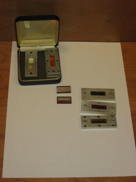
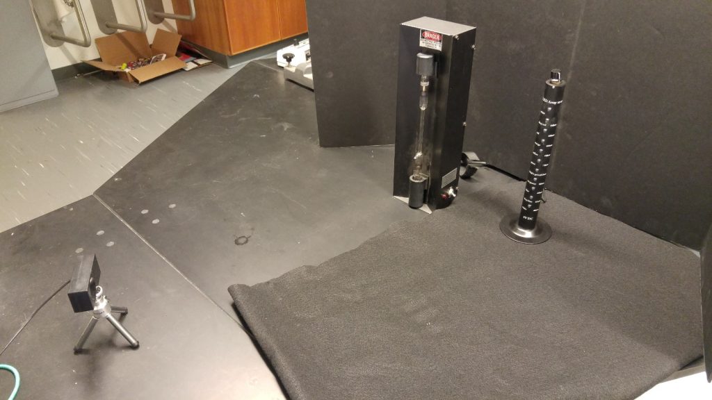
- The LED tower (shown in top photo above, next to spectral tube) contains 11 different LED’s of various wavelength, and pairs well with the camera as a demo.
- Located in L35, Isle G, top shelf, next to diffraction gratings.
light
Laser light bend
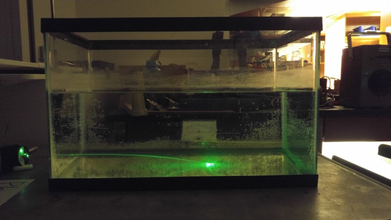
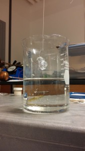
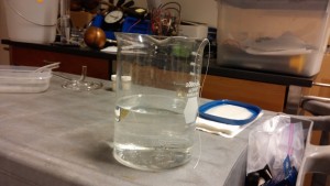
- Tank shown above contains a solution of sugar water that becomes more and more concentrated with depth. As sugar water has a higher index of refraction than pure water, the sugar density gradient produces a depth-dependent index of refraction. A laser beam shot through the solution, initially parallel to the bottom of the tank, will bend downward and hit the bottom.
- To prepare solution: pour sugar into empty tank, a few centimeters thick. Then, using a hose, very slowly (so as not to agitate sugar) fill the tank with water about halfway full.
- Tank located in L01, section B6. Sugar in L35, under sink. Laser in L35, section A5.
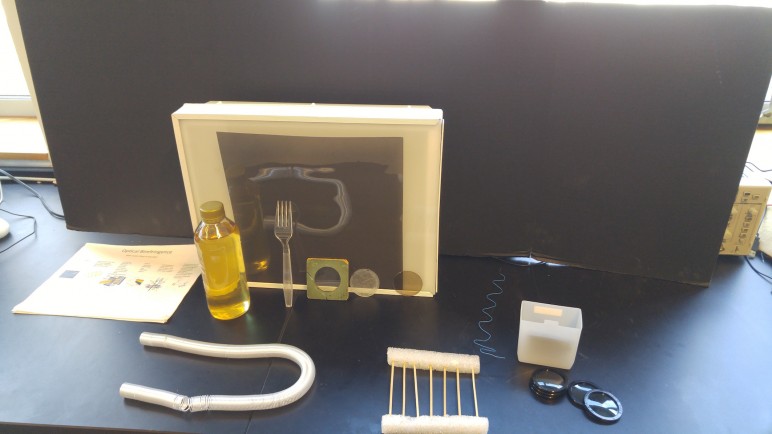
Polarization (outreach set)
Demos in this set include:
- Planar light source with large polarizing sheet
- Corn syrup
- Long spring
- Plastic fork
- Wafers of micah and benzoic acid crystals
- Cubes of calcite, selenite, and Iceland spar
- Disappearing cube
- Polarizer model (looks like a gate), with light model (wavy wire piece)
- Square and circular polarizers
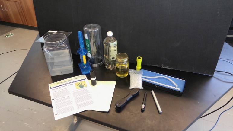
Light and Lasers (outreach set)
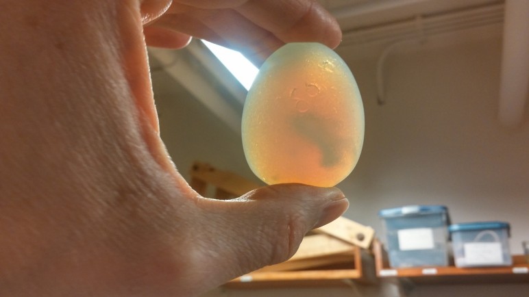
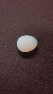
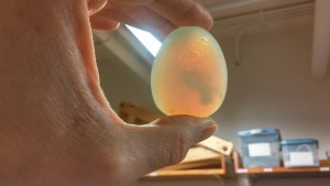
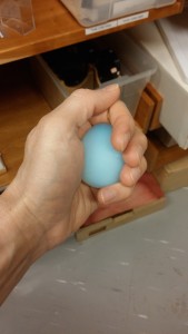
Sunset Egg
Appearance of egg mimics that of the sky.
Glass egg is translucent, milky white, and contains no pigment- only microscopic particles that scatter light. Short wavelengths of light (blue) have a tendency to scatter off the microscopic particles in the glass; while long wavelengths of light (red and yellow) tend to pass through. The egg therefore appears yellowish red when held in front of a light source, and blue when held in your hand.
- Located in L01, section B-6
Past Light Cone (to Big Bang)
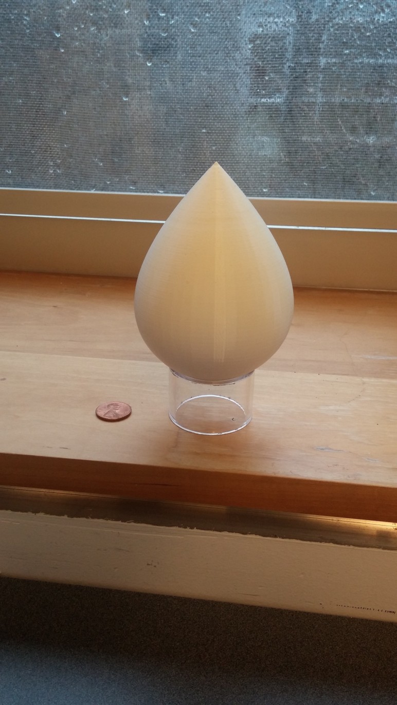
In a Big Bang universe, the shape of our past light cone is not strictly conical, but tear-drop shaped. The moment of now is located at the pinnacle of the drop, the moment of the Big-Bang is located at the very base.
To understand why our past light cone has this shape, recall that the distance from a light cone’s surface to its time axis is the photon’s emission distance. All of the photons we see today- from the very early universe- were emitted from regions of space that were, at the time, very close to us. (Spatial expansion caused these regions to separate very rapidly.) So, photons emitted very early in the universe’s history, and photons emitted very recently have small emission distances.
The slope of the light cone represents the recessional velocity of light with respect to our region of space. The fattest part of the light cone corresponds to the moment in time when the photons we are currently seeing, from the Big Bang, first began to approach us.
The model shown above is actually the light cone for a linearly expanding universe (expansion at a constant rate- i.e., recessional velocities do not change with time). The most accurate model for spatial expansion- lambda cdm- is actually very close to this in shape.
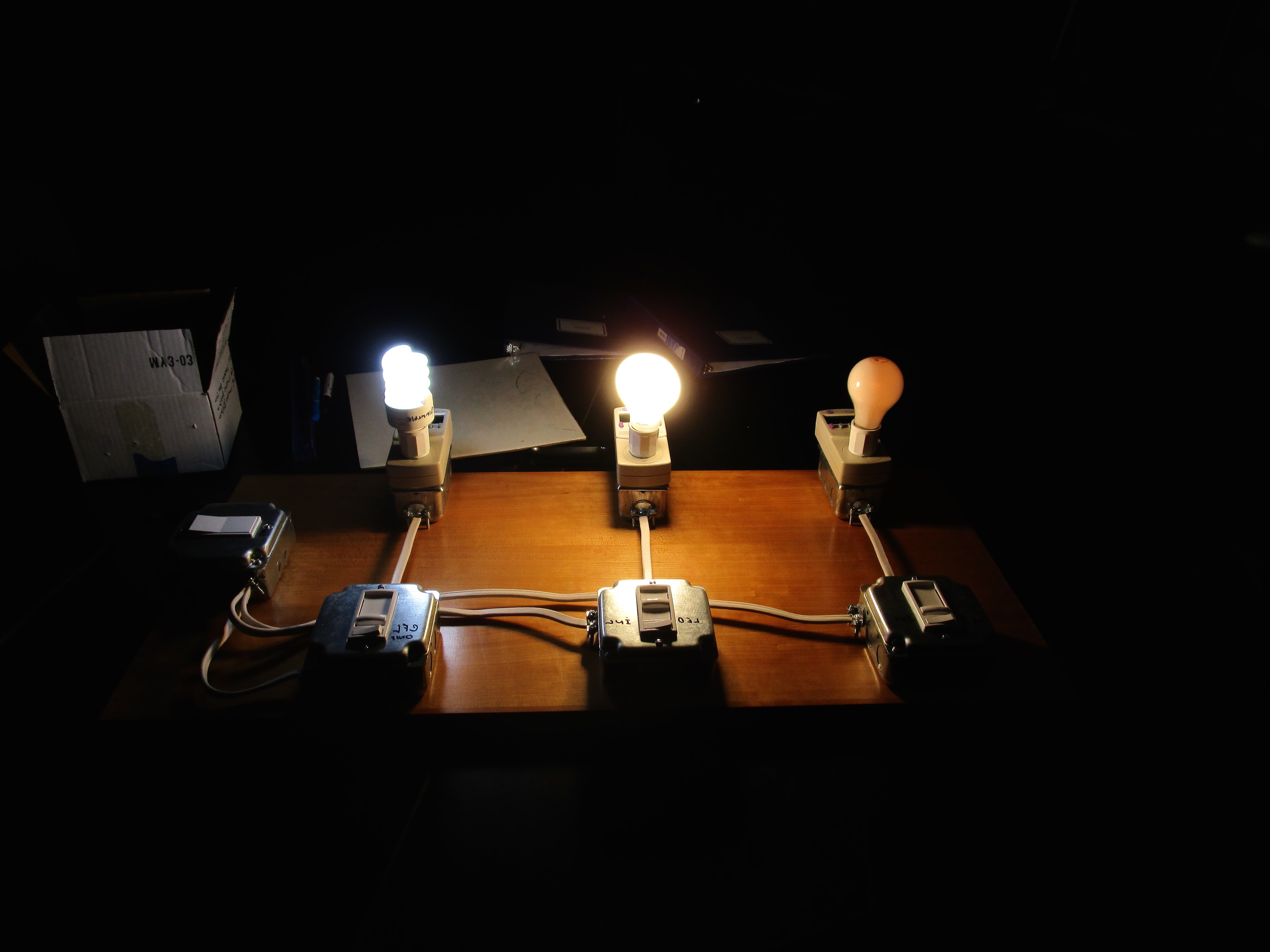
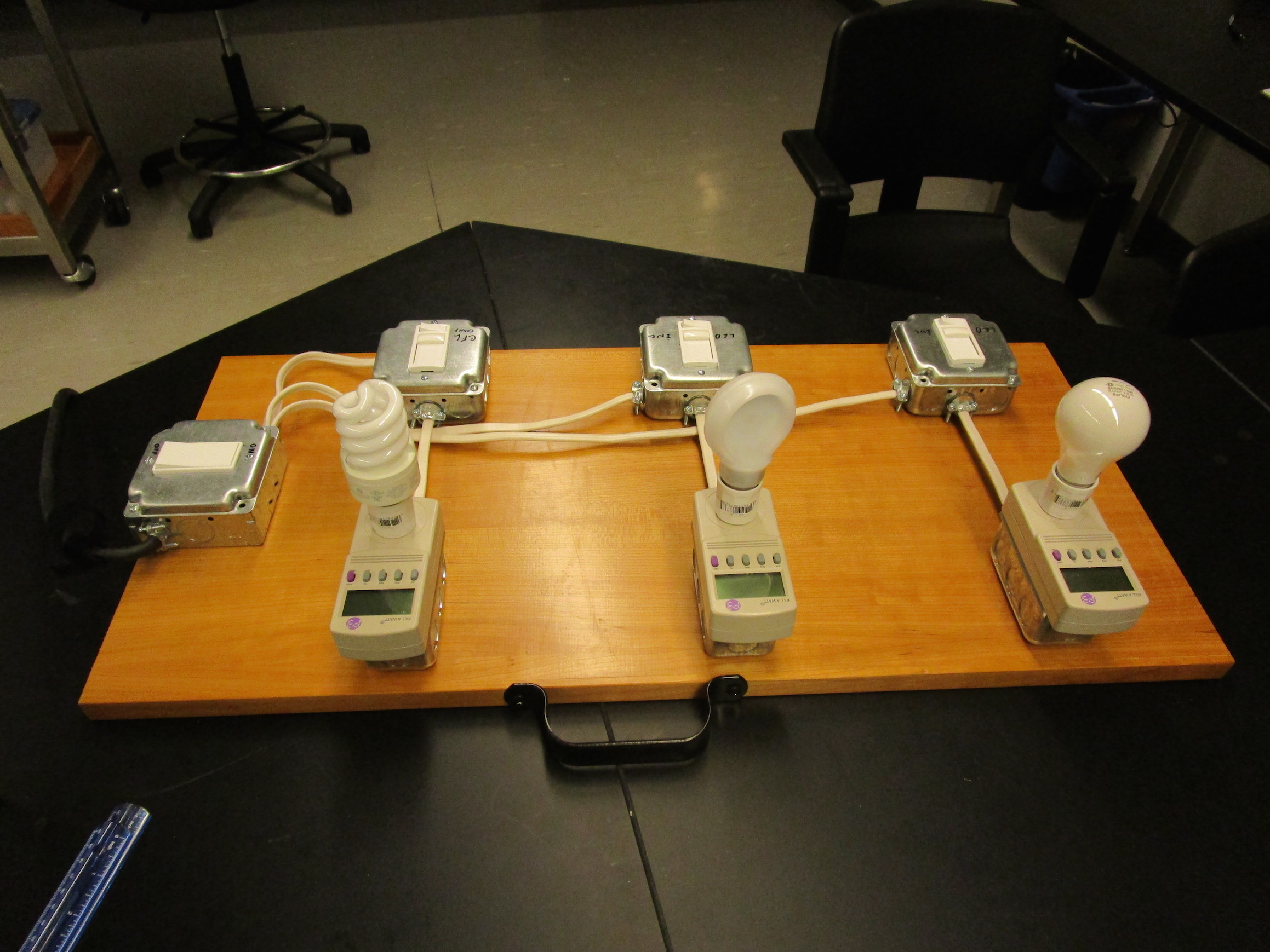
Light Bulb Energy Usage
- When bulbs consume the same amount of power (same wattage) their brightness differs substantially- the incandescent bulb is much dimmer than both the compact fluorescent and the LED, which are of comparable brightness.
- Bulb power can be adjusted using sliders. Meter connected to bulb tells both watts and volts.
- Located in L01, section B5
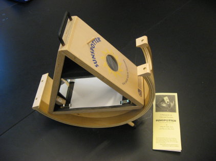
Sunspotter
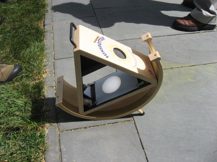
- Use to see sunspots.
- Projects a 3″ image of the sun onto a white viewing screen. Very easy to set up. Instructions are included.
- Located in 187.
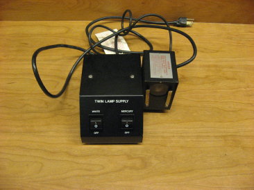
White light / Mercury lamp
- Located in L01, section C1.
- Danger: Even momentary exposure to ultra violet rays
causes severe eye damage. Always keep the opening of the light box covered
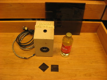
with one or more glass plates or filters when unit is used as light source. Never look directly at the Mercury Vapour lamp if
the glass plate is removed.
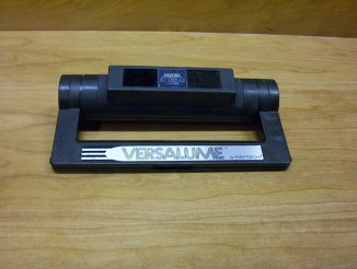
Blacklight
- Use to illuminate mineral samples.
- Caution: Do not expose your eyes to the light
from this lamp. - Located in L01, section C1.
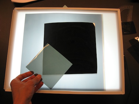
Crossed Polarizers
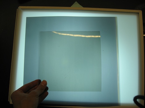
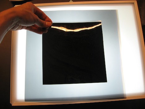
- Light is totally blocked when two polarizers are oriented 90 degrees apart. When third polarizer is inserted between the first two at an intermediate angle, light is transmitted.
- Located in L01, section B-5.
Optical Rotation Bowl
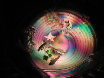
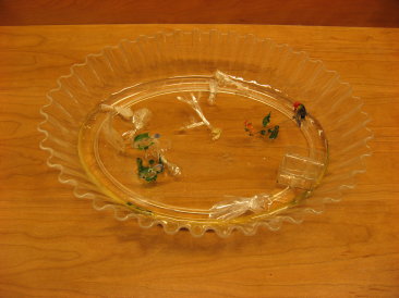
- Illuminate bowl with polarized light. Bowl, and objects within,
appear multi-colored when viewed through a polarizer. Rotate polarizer to
see colors change. - Phenomenon is the result of optical rotation. Linearly polarized light gets rotated by molecular composition of bowl; extent of rotation
depends on wavelength of light, causing angular separation of colors. - Located in L01, section B5.
Barber Pole
- Shine polarized light through a bottle of corn syrup. Bottle of syrup appears multi-colored when viewed through a polarizer. Rotate polarizer to see colors change.
- Phenomenon is the result of optical rotation. Syrup rotates linearly polarized light; extent of rotation depends on wavelength of light, causing angular seperation of colors.
- Located in L01, section B5.
Scattering and Absorption
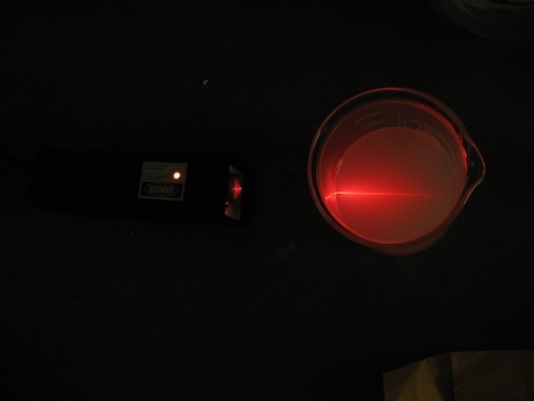
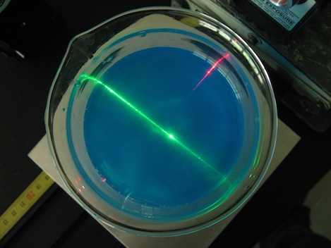
- Red laser light penetrates milky solution and is slightly attenuated (top photo).
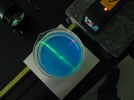
- Blue dye absorbs red light but not green light. Red laser attenuation is much greater than green in blue-dye/water solution.
Location
- Lasers: L35, section A-5.
- Dye: L35, in cabinet above sink.
- Beaker: L35, section G-3.
Spectroscopes
- Located in L01, section C1.
Interference Filters
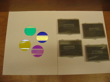
- Glass plates coated with different thicknesses of dielectric
material transmit and reflect different wavelengths of light. - Located in L01, section B5.
Phantom Crystals
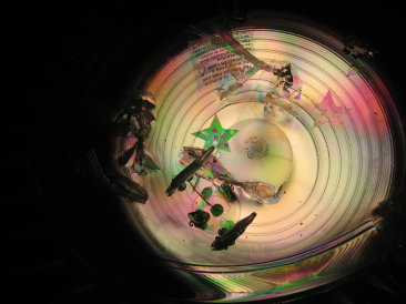
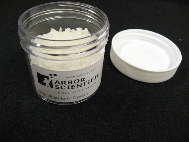
- Phantom Crystals are carbon-based polymers that absorb up to 300 times their weight in water. A fully saturated crystal in a glass of water is almost invisible, as light passes through it without being refracted. But when exposed to air, the water soaked crystals are clearly visible, because air’s index of refraction is very different from that of water.
- Located in L01, section B-5
Atomic Spectra
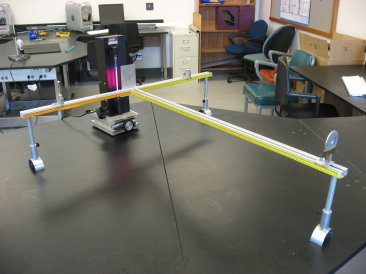
- View Balmer series using hydrogen light source, diffraction
grating, and optics rail.
Location
- Optics rail parts: L02, section B6
- Spectrometer grating (small one, in above photo): L35, section A5
- Spectrometer grating (large, hand-held, good for demos): L02, section C1
- Spectral tubes and power supply: L35, section G1
Diffraction of light
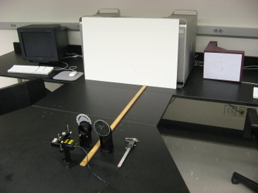
- Shine laser beam through a variety of diffraction patterns
to demonstrate properties of single and multiple-slit diffraction. - Diffraction Accessories located in
L01, section B5. - Calipers located in L35, section D2.
1/d^2 Dependence
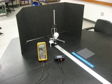
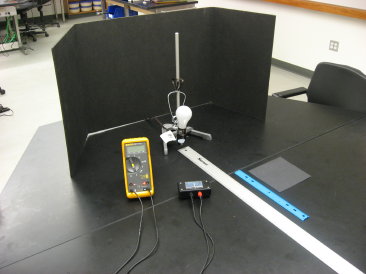
- Purpose:Demonstrate 1/d^2 dependence of brightness.
- Adjust distance of light detector, current meter indicates
brightness of light.
Location
- light detector: L35, section G1
- light bulb, socket, and clamp: L35, section D2
- ring stand: L35, section A4
- 2 meter stick: L35, section A2
Human Eye Model
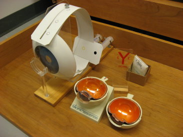
- Located in L01, section B6.
Blackboard Optics
Experiments Include
- The laws of reflection
- virtual image with a plane mirror, convex mirror, and a concave lens
- focal length of concave and convex mirrors
- focal length of planoconvex and planoconcave lenses
- real image formed by a concave mirror
- simple refraction
- less dense to more dense medium
- parallel displacement by a rectangular block
- semicircular body
- light incident at center if disc and at right angles to tangent
- critical angle
- total internal reflection
- reversing prism.
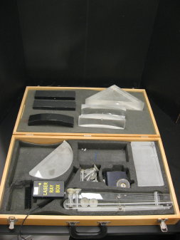
Have 2 sets. New set uses magnetic attachments, old set uses suction.
- Located in L01; section B6